Treatment of external iliac artery stenosis using lower profile 9 mm GORE® VIABAHN® Endoprosthesis

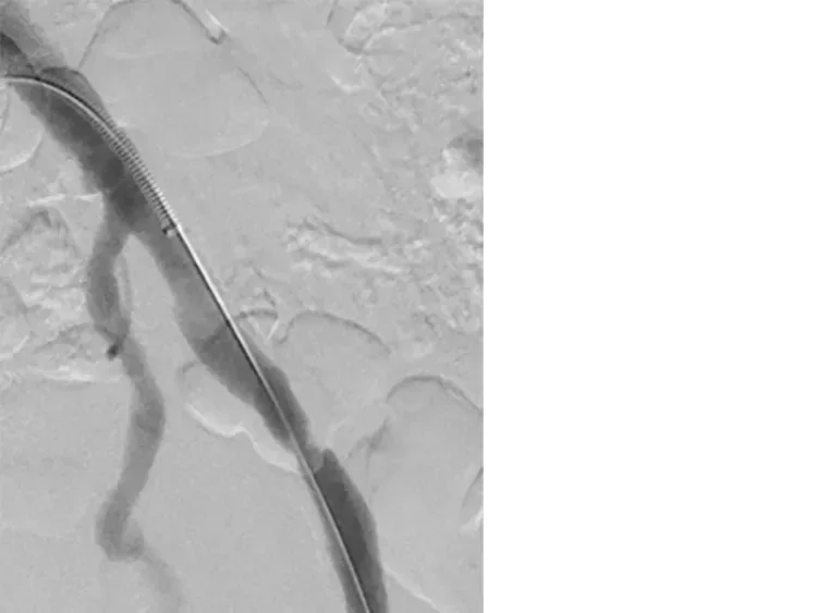
Angiogram with sheath in left common iliac artery demonstrates severe stenosis within mid external iliac artery
Challenge
70-year-old male with painful dry gangrenous left second toe, present for two months. No evidence of cellulitis or wet gangrene, however, the patient was treated with antibiotics for two weeks prior to the procedure.
- Associated ischemic rest pain in the toe that improved with dependent positioning confirmed by duplex ultrasound with monophasic waveforms throughout the lower extremity (Ankle-Brachial-Index (ABI) 0.87 and Toe-Brachial-Index (TBI) 0.0). No other symptoms of rest pain or claudication in the foot or leg.
- Relevant patient history:
- Diabetes mellitus type II, hypertension, chronic kidney disease type III, hypocalcemia.
- In remission from seminoma after a left radical orchiectomy and left pelvic primary radiotherapy, which the physician suspected may have resulted in embolizing iliac artery lesion.

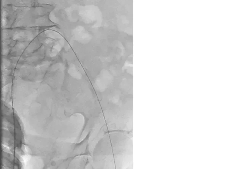
Image: Up-and-over the aortic bifurcation, the GORE® VIABAHN® Endoprosthesis with PROPATEN Bioactive Surface was placed within the left external iliac artery, prior to post-dilation
Image courtesy of Charles Briggs, M.D. Used with permission.
Procedure
The procedure plan was to minimize contrast dye by using intravascular ultrasonography (IVUS) to evaluate for left iliofemoral embolizing and/or flow-limiting lesions and perform an angiogram of the popliteal-tibial runoff to assess healing potential of toe and need for distal revascularization.
- Right femoral access was gained with a 6 Fr sheath and then an ANGIODYNAMICS® OMNI Flush Angiographic Catheter and TERUMO® RADIFOCUS® GLIDEWIRE® ADVANTAGE Guidewire were advanced up-and-over the aortic bifurcation.
- The wire and straight-tip catheter were advanced into the left popliteal artery and a runoff angiogram was performed with no treatable tibial arterial lesion identified.
- The TERUMO® RADIFOCUS® GLIDEWIRE® ADVANTAGE Guidewire was exchanged for a .014" ABBOTT® HI-TORQUE SPARTACORE Guidewire into the left popliteal artery. A .014" PHILIPS EAGLE EYE PLATINUM RX Digital IVUS Catheter was advanced distally over the wire, where severe stenosis within the left external iliac artery and left popliteal artery were identified.
- The wire was exchanged back to a TERUMO® RADIFOCUS® GLIDEWIRE® ADVANTAGE Guidewire and a 6 Fr TERUMO® Sheath was advanced over the wire to left common femoral artery (CFA).
- The popliteal artery was ballooned with a 5 x 120 mm BOSTON SCIENTIFIC MUSTANG Balloon Dilatation Catheter, followed by a 6 x 150 mm MEDTRONIC IN.PACT Admiral DCB. Completion angiogram demonstrated satisfactory resolution of stenosis.
- The up-and-over sheath was then exchanged for an 8 Fr TERUMO® Sheath.
- The sheath was then pulled backed into the left common iliac artery (CIA), and IVUS identified stenosis in the external iliac artery (EIA).
- Diameter measurements were taken at the lesion, also proximal and distal to the lesion for stent sizing (vessel measured 8.5 mm).
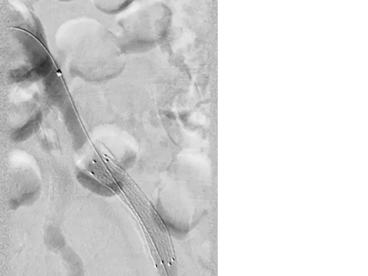
- A 9 mm x 5 cm GORE® VIABAHN® Endoprosthesis with PROPATEN Bioactive Surface was selected and deployed across the lesion.
- Post-dilation was performed with a BOSTON SCIENTIFIC MUSTANG Balloon Dilatation Catheter.

Image: Resolution of left EIA stenosis using a 9 mm x 5 cm GORE® VIABAHN® Endoprosthesis with PROPATEN Bioactive Surface
Image courtesy of Charles Briggs, M.D. Used with permission.
Result
- Completion IVUS and angiogram showed good expansion of the VIABAHN® Device without residual stenosis.
- Patient was discharged same day on acetylsalicylic acid and clopidogrel.
- Follow-up in clinic in thirty days revealed healing of the left second toe.
- Plethysmography revealed left ABI increase to 0.98 with associated increase in TBI to 0.50.
- Arterial duplex ultrasonography showed the treated area to have triphasic flow in the external iliac artery and infrainguinal arterial tree.
Case Takeaways
- For this patient, extensive IVUS was helpful to identify the lesion, normal lumen proximal and distal to the lesion, and precise measurements. IVUS was also instrumental in accurately measuring the vessel diameter and selecting the appropriate VIABAHN® Device diameter per the sizing table in the IFU.
- The ability to deliver the VIABAHN® Device through an 8 Fr sheath greatly improves the ability of the interventionalist to safely treat larger caliber vessels with a lower likelihood of access site complications.
- The use of covered stents protects from possible extravasation in highly calcific vessels and may perform better in more complex anatomy.1 Since there is a fair bit of tortuosity within the mid- to distal- external iliac artery, the flexibility of the VIABAHN® Device may be more suitable at managing this anatomy.
*As used by Gore, PROPATEN Bioactive Surface refers to Gore’s proprietary CBAS Heparin Surface.
1. Lammer J, Dake MD, Bleyn J, et al. Peripheral arterial obstruction: prospective study of treatment with a transluminally placed self-expanding stent graft. Radiology 2000;217(1):95-104.
ABBOTT and HI-TORQUE SPARTACORE are trademarks of Abbott Laboratories. ANGIODYNAMICSR and OMNI are trademarks of AngioDynamics, Inc.
BOSTON SCIENTIFIC and MUSTANG are trademarks of Boston Scientific Corporation.
MEDTRONIC and IN.PACT are trademarks of Medtronic, Inc.
QUICK-CROSS is a trademark of Spectranetics Corp.
TERUMO, RADIFOCUS and GLIDEWIRE are trademarks of Terumo Medical Corporation.
This content is for informational purposes only, is not advice or a guarantee of outcome. It is not a substitute for professional medical advice, diagnosis or treatment. Individual results and/or treatment may vary based upon the circumstances, the patient’s specific situation, and the healthcare provider’s medical judgment.
Refer to Instructions for Use at eifu.goremedical.com for a complete description of all applicable indications, warnings, precautions and contraindications for the markets where this product is available.